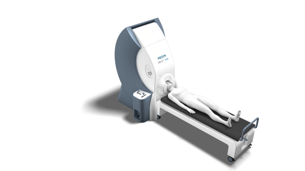
Using MEG alongside other modalities
A comparison
fMRI is used to localize brain functions prior to surgery. This offers an indirect measure of brain activity with poor temporal resolution.

MEG is a direct measure of electrophysiological activity within the brain and may therefore more accurately detect actual brain activity.
Long-term monitoring by EEG requires large numbers of electrodes to be consistently positioned on the subject’s head. Localization accuracy is poor due to the conduction of the signal through the skull and the scalp.

Greater accuracy of source localization is possible with MEG as the skull and scalp are transparent to the magnetic signals, allowing a consistent, clean signal. Propagation of epileptic activity from one area of the brain to another can be monitored with MEG.
SPECT is highly invasive. It requires a contrast medium to be injected and the patient to be having an epileptic seizure.

MEG is non-invasive. The patient experience is peaceful and comfortable. There is no need to inject contrast agents or require patients to undergo epileptic seizures. Many patients fall asleep during their MEG scan.
Intracranial EEG is an accurate technique for localizing and confirming epileptic areas. However, it requires brain surgery and has limited spatial coverage and resolution.

MEG does not require placing invasive electrodes. MEG can be used to monitor most brain areas completely non-invasively.
Using MEG alongside other modalities
A comparison
fMRI is used to localize brain functions prior to surgery. This offers an indirect measure of brain activity with poor temporal resolution.

MEG is a direct measure of electrophysiological activity within the brain and may therefore more accurately detect actual brain activity.
Long-term monitoring by EEG requires large numbers of electrodes to be consistently positioned on the subject’s head. Localization accuracy is poor due to the conduction of the signal through the skull and the scalp.

Greater accuracy of source localization is possible with MEG as the skull and scalp are transparent to the magnetic signals, allowing a consistent, clean signal. Propagation of epileptic activity from one area of the brain to another can be monitored with MEG.
SPECT is highly invasive. It requires a contrast medium to be injected and the patient to be having an epileptic seizure.

MEG is non-invasive. The patient experience is peaceful and comfortable. There is no need to inject contrast agents or require patients to undergo epileptic seizures. Many patients fall asleep during their MEG scan.
Intracranial EEG is an accurate technique for localizing and confirming epileptic areas. However, it requires brain surgery and has limited spatial coverage and resolution.

MEG does not require placing invasive electrodes. MEG can be used to monitor most brain areas completely non-invasively.
fMRI vs MEG
Functional magnetic resonance imaging or functional MRI (fMRI) measures brain activity by detecting changes associated with blood flow within the brain. This technique is predominantly used to localize brain functions prior to surgery.
The difference between these two techniques predominantly lies in that fMRI measures blood flow relying on the fact that cerebral blood flow and neuronal activation are coupled. MEG directly measures brain activity through the magnetic field the neuronal activation produces. Consequently, MEG has a much higher temporal resolution than fMRI, allowing the timing of brain activity to be much more precisely measured.
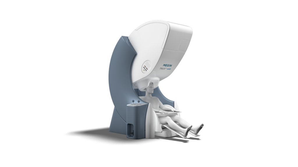
EEG vs MEG
Similar to MEG, EEG measures and records electrical activity in the brain. However, unlike EEG, MEG can tell where in the brain this functional activity originates. This is because the human skull and the tissue surrounding the brain distort the electrical signals an EEG picks up, but affects magnetic fields produced by those electric signals much less and in a measurable way that can be accounted for.
EEG and MEG are great tools to use in conjunction with one another as the information they offer complement one another.
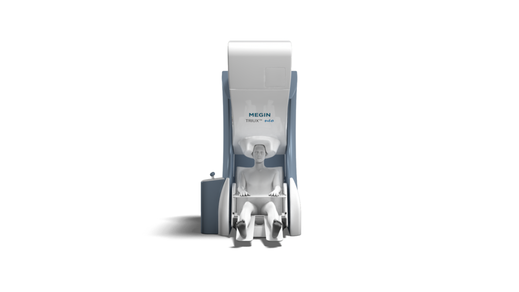
SPECT and PET vs MEG
A single photon emission computed tomography (SPECT) scan and a positron emission tomography (PET) are imaging modalities that show how blood flows to tissues and organs. Both are nuclear imaging scans that integrates computed tomography (CT) and a radioactive tracer. The tracer is what allows doctors to see how blood flows to tissues and organs. Before the scan, a tracer is injected into the bloodstream. This imaging technique is used to form a 3D image of the brain. MEG is completely non-invasive and does not require any tracer, radioactive or otherwise, to be injected.
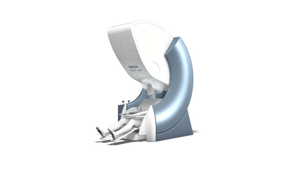
iEEG vs MEG
Intracranial electroencephalography (iEEG) is a technique that measures and records electrical brain activity just as scalp EEG does. However, iEEG rquires electrodes to be implanted in the brain. This allows it to be much more accurate with respect to the location and timing of electrical activity in the brain. However, this method requires brain surgery. And because of this, iEEG cannot be used for all brain areas due to difficulty of access to all parts of the brain. MEG allows for accurate measurement of the location and timing of electrical activity in the brain, in a quick and easy non-invasive scan.
MEG vs CT
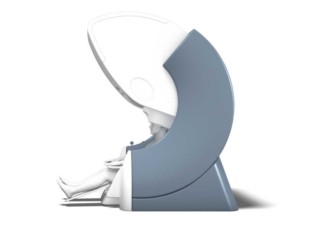
A computerized tomography (CT) scan combines a series of X-ray images taken from different angles around your body and uses computer processing to create cross-sectional images of the bones, blood vessels and soft tissues inside your body. A CT scan is a standard imaging modality used to assess the brain. However, because it only creates a structural image of the brain, it is not an imaging technique that can determine how the brain is functioning. In addition, CT scans use ionizing radiation which has given rise to concern about radiation exposure in patients who have had many scans. MEG is completely non-invasive and does not require the use of radiation to detect brain function.
MEG vs MRI
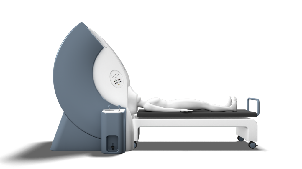
Magnetic resonance imaging (MRI) is a medical imaging technique used to form pictures of the anatomy and the physiological processes of the body. MRI scanners use strong magnetic fields, magnetic field gradients, and radio waves to generate images of the organs in the body. Because of it’s use of magnets to image the body, some patients with certain types of implants are not able to have an MRI scan. However similar to CT, MRIs creates a structural image of the brain and therefore isn’t an imaging technique that can asses how the brain is functioning.
